Figure 1. PDGF BB binding sites determined by C Finder. Critical amino acid residues in binding receptor in subunit I (A) and II (B) of native PDGF, 3D structure of native PDGF BB (C), and 3D structure of single-chain PDGF (D). The binding sites candidate to be modified, substituted amino acid residues, and the linker are shown in red, green and yellow, respectively.
2025-01-22 | | |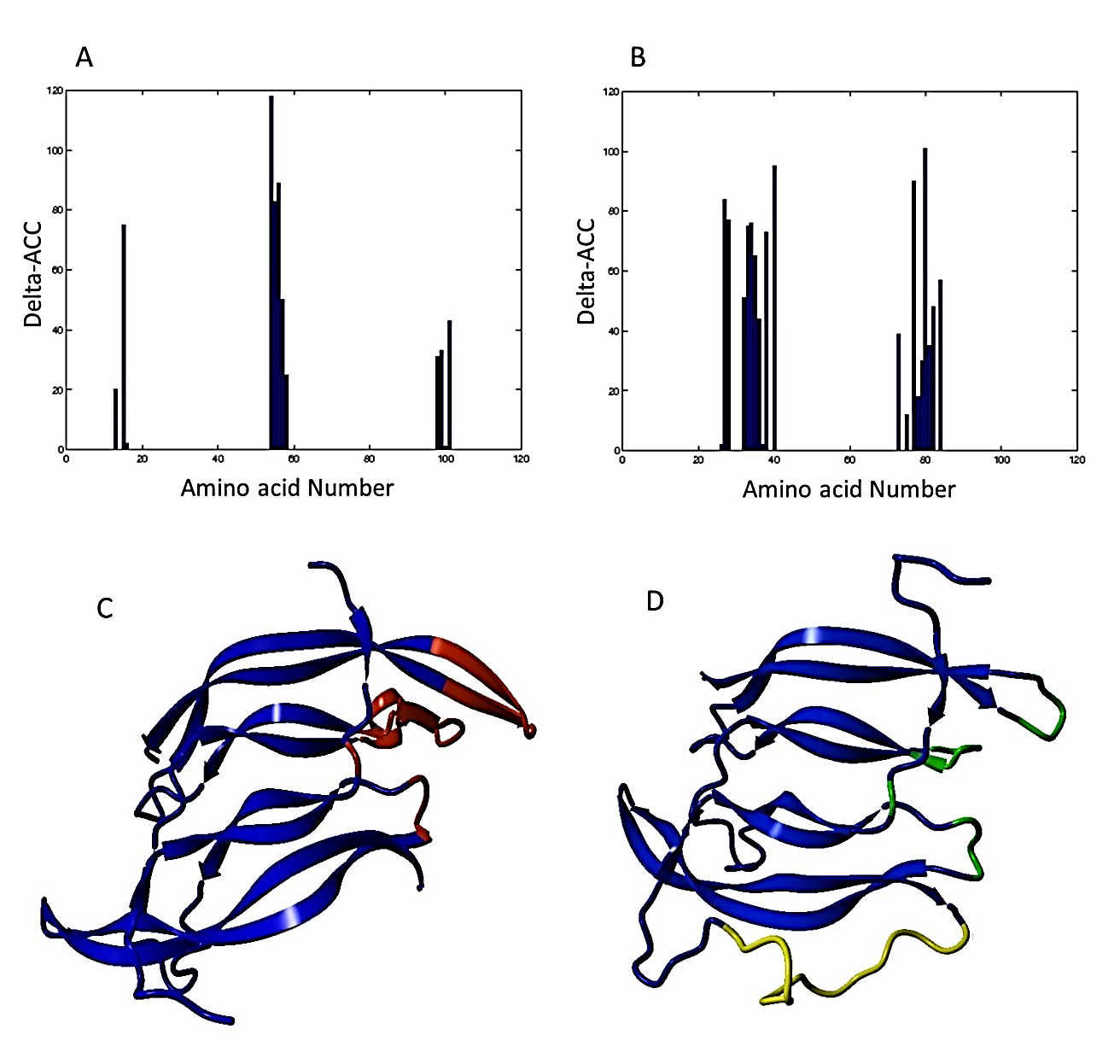
Figure 1. PDGF BB binding sites determined by C Finder. Critical amino acid residues in binding receptor in subunit I (A) and II (B) of native PDGF, 3D structure of native PDGF BB (C), and 3D structure of single-chain PDGF (D). The binding sites candidate to be modified, substituted amino acid residues, and the linker are shown in red, green and yellow, respectively.
(ISSN - Online)
2959-8591