INTRODUCTION
Reinforced concrete is considered one of the essential materials used in structures due to its durability, longevity, and load-bearing capacity. However, the long-term exposure to various environmental factors, especially in coastal areas or those exposed to salts, leads to the corrosion of the reinforcing steel (1). This corrosion produces iron oxides in volumes far greater than those of the original reinforcing steel. This causes internal tensile stresses in the surrounding concrete, which eventually manifest as surface cracks. These cracks typically follow specific patterns (such as cracks parallel to the steel bars or branching cracks), reflecting an advanced stage of corrosion. Traditional methods for examining these cracks often rely on visual inspection and direct geometric measurements, but these methods may not capture the complexity of their propagation patterns and the rate of their development.
Hence the importance of Fractal Geometry as a mathematical tool capable of describing the complexities of irregular and intricate shapes in nature, which are difficult to describe using traditional Euclidean geometry. This research proposes a fractal body that corresponds to the shapes of cracks taken from images of damaged structures, then applying digital image processing techniques to extract cracks from images of concrete cracks, and finally analyzing them quantitatively using the Fractal Dimension, which reflects the complexity and branching of the cracks. In this research, the Box-Counting method was applied at different scales and the results were compared with the proposed fractal body. Wang (2012) and others quantitatively determined the surface cracking pattern in AAR (alkali-aggregate reactant concrete) using fractal geometry. This paper presents a novel evaluation method based on AAR crack analysis. The cracking pattern on the damaged surface was determined using image analysis. The results indicate the conditions for fractal analysis, fractal properties, and classification of AAR cracks (2). Zaborac (2019) and other researchers applied two methods, one mechanical, to estimate shear strength in the presence of diagonal cracks, and the other is to perform a fractal analysis by processing crack images, with the aim of improving methods for evaluating cracks in concrete within bridges. The results showed that crack width alone is an insufficient indicator to predict the severity of the damage, and it is necessary to combine the engineering data of the crack with modeling to provide a more accurate assessment. The method based on fractal analysis also allowed for the differentiation of different levels of damage (light, medium, severe), making it a helpful tool for engineers to estimate the condition of bridges more quickly and accurately (3). Ji (2020) and other researchers proposed a quantitative index that describes the degree of internal corrosion expansion in reinforced concrete using fractal geometry. This approach allows for the representation of similarity and complexity in the development of cracks resulting from corrosion in concrete. They studied the effect of the cracking pattern and the distribution of coarse aggregate on the distribution of cracks, and used the partial immersion galvanic corrosion acceleration method to obtain the distribution of cracks within the elements. The results showed that the cracking pattern was the main factor affecting the complexity of crack distribution; in cracks with the simplest cracking patterns, the presence of coarse aggregate and the irregularity of its surfaces strongly affect the direction of crack growth (4). In their research paper, Khan (2023) and other researchers reviewed various image processing techniques used to detect cracks in concrete, along with a scientific analysis of previous research. The study included traditional methods such as borderline detection, thresholding and noise filtering, as well as modern approaches using artificial intelligence and deep learning (5). The study did not apply fractal analysis itself, but through it; it was possible to identify auxiliary processing techniques for detecting cracks and processing images. Thybo›s (2018) research focused on simulating the propagation of corrosion in reinforcing steel and its consequences on the surrounding reinforced concrete. The model used was divided into five basic zones: concrete, reinforcement, corrosion layer, cracking, and adhesion separation at the interface between steel and concrete. The researcher imposed a hypothetical thermal load on the corrosion layer that simulates the swelling of rust products, and used a cracking model to simulate crack opening and movement at the reinforcement surface. The research aimed to improve service life models for reinforced concrete in cases where corrosion occurs (6). The research did not directly use fractal analysis, but it provides important background on the relationship between corrosion and structural cracking. The proposed model also demonstrated that cracks are not only superficial but are related to corrosion in steel and propagation within concrete, which supports the hypotheses that intend to use fractal dimension as an evaluation mechanism. An (2022) and other researchers proposed a new method that combines fractal dimensionality and a UHK-Net (a neural network used in image processing) for the semantic recognition of cracks in concrete. The research relies on calculating the local segmentation dimension to determine the possible locations of the cracks; then the neural network is used to accurately segment the image (7). The fractal dimension was used as an additional indicator in the processing stage. Cheng (2023) and other researchers presented an algorithm for detecting cracks, segmenting them, and estimating the fractal dimension in low-light conditions by combining the Fourier transform with a neural network to improve segmentation in dark images. The algorithm improves the discrimination between noise and the true lines of the crack, and then applies fractal calculations to the extracted lines (8). The fractal dimension was actually applied after segmentation in low-light conditions.Wang (2025) discussed the problem of choosing scale criteria and starting count when applying the Box-Counting method, as random choices may affect the stability and comparability between studies, and this restricts the actual engineering application of the method (9). Researcher Xie (2024) proposed a method for assessing damage in concrete components that relates the U-Net and calculates the fractal dimension. A linear regression equation was then constructed between the fractal dimension and the damage coefficient. The researcher tested the method on a sample of laboratory concrete wall and found that the classification accuracy was about 83.33% using this method (10). Ai (2023) presented a modified method for calculating fractal dimension using square counting in a more direct way to reduce errors. His research suggests that some counting points are modified or deleted to reduce bias in the higher ranges of accuracy (11). Li (2022) and other researchers conducted an experimental study of the relationship between surface cracks of concrete surfaces and the degree of corrosion of steel bars using fractal theory. Samples were prepared with reinforcement of smooth HPB300 reinforcing steel according to standard dimensions, which vary in both the diameters of the steel bars and the rate of corrosion. The steel bars in the structural frameworks were partially immersed in a saline solution to ensure that the electrolyte solution was in sufficient contact with the surface of the steel bars. The semi-immersion method was used in this study because the corrosion of the steel bar should occur as in the natural environment. The concrete samples were semi-immersed in a 5% sodium chloride solution to ensure that the solution penetrated through the capillary permeability of the pores in the concrete into the steel. The fractal dimension reflects the space occupied by the nodal shapes, and is a measure of the irregularity of these shapes (12). The study was based on two methods: The Box-Counting Method, also known as the Covering Algorithm, which relies on covering the
fractal curve with square boxes of different sizes. The measurement of the fractal dimension is based on measuring the slope of the surface roughness only (13). The Pixel-Covering Method: A digital image is stored as pixels, and a high number of n pixels is represented as an array (n x m) where each element in the array represents a pixel. It is converted into a grayscale image using MATLAB (14). Angel (2014) studied the fractal effect of corrosion on the mechanical behavior of unprotected A36 steel exposed to corrosion (steel that has not been coated with any coating techniques using zinc as the primary protective element), which leads to the observation of cracks in the steel in marine environments. The research is based on analyzing the dimensions of the samples, conducting chemical laboratory analyses, and finally conducting a fractal analysis of the tested samples (15). Yao (2019) and other researchers conducted a study of fractal models of concrete cracks exposed to sulfate attack. They immersed concrete samples in an 8% sodium sulfate solution for one day, then removed and dried them. Corrosion tests were then carried out one month after the samples had been stored in nylon bags, and the rates of surface crack propagation were calculated and evaluated at different corrosion time points. It was concluded that although the surface cracks are complex and spread in all directions, they can be described by the fractal dimension, which is an exponential function of time. The fractal dimension doubles with increasing erosion time according to the rate of chemical reaction. The higher the water-to-cement ratio in the concrete, the greater the degree of damage and the fractal dimensions of the samples (16).
After reviewing numerous reference studies and presenting a selection of them, it became clear that most previous research neglected to address the root causes of crack problems. Some of the proposed solutions that may be inadequate for the current situation. These solutions were experimental and, when applied in practice, failed relatively to consider crack types, weathering and erosion factors, the geological characteristics of the terrain, the slowness of implementation, and other factors. Other solutions included surface treatments (carbon fiber filling, steel reinforcement, etc.), and some research suggested preventative measures. Logistical challenges were also highlighted, such as the difficulty of accurately predicting the crack›s location and position, accurately diagnosing its nature and cause, and monitoring its development (before and after treatment) over an extended period. On the other hand, computer simulation technology has emerged as a promising solution for addressing such problems in building cracks. Despite advancements in simulation and the development of codes to address crack defects and deficiencies, some proposed applied studies have lacked a formula for representing true cracking and have encountered problems in simulating the interaction between the soil, foundations, and structure. Some of these studies rely on only one indicator (crack width) in addition to the fractal dimension to analyze concrete crack images. Among the modern solutions proposed—which this research aims to implement—is the use of digital images and their computer simulation. The study presented here is based on the observation of a recurring fractal structure in formed concrete cracks. We proposed a simple shape for this structure to represent the crack, then used the box counting method to compare it to this fractal structure and calculate several indicators that describe the cracks more accurately. Therefore, the objective of this research is to evaluate concrete cracks using a computer program that defines both the structural structure and its indicators, and generates a code for analyzing crack images. On the other hand, this research is significant in saving time by rapidly assessing the extent of damage caused by concrete cracks, thus reducing the cost of traditional structural evaluation methods. Furthermore, its novelty lies in its creation of an initial research foundation for developing other solutions, including experimental applications combining nanotechnology with computer simulations, to provide effective and robust solutions for various types of crack problems and defects.
MATERIALS AND METHODS
The methodology was divided into several stages. Initially, images of cracks in various concrete elements of buildings in the Latakia Governorate were collected to create a database containing the stored images along with their metadata. Ten photos were taken for each crack location, and these photos were evaluated to determine which were the most accurate in terms of lighting, brightness, visibility of internal damage, and noise isolation. Then, the fractal body was identified, and the necessary calculations were performed to monitor crack behavior and assess crack severity. After that, the images were enhanced by adjusting brightness and removing noise.
Then, the box-counting method was applied, relying on converting the real image to a binary (black and white) image, and then performing the following steps:
a) Choosing box sizes: by selecting several square sizes that are multiples of 2, Ɛ={2,4,8,16,32,64} in pixels.
b) Covering the crack image with a grid of squares, then counting the squares that contain at least one pixel of the crack. This count is N(Ɛ). To ensure accurate results, only the path of the crack was covered with squares, leaving the rest of the image uncovered.
c) Converting the values to logarithms, where: Xk=log(1/Ɛk) and Yk=log(N(Ɛk)) to draw a log-log plot, meaning that each point in the plot is a k(Xk,Yk).
Finally, aset.of.indicator.was.culated.to.evaluatethecracks:f.indicator.was.culated.to.evaluatethecracks:
The fractal dimension, which represents the degree of irregularity and complexity of the crack path, can be calculated using the following formula:
 Where:
Ɛ: the side length of the square in the grid, N(Ɛ): the number of squares through which the crack passes, and D: the fractal dimension of the crack path.
If D≈1, the crack is linear; conversely, the higher the value of D, the more complex and rough the crack becomes
II. The degree of branching, which represents the density of branches in a crack, can be calculated using the formula: F=Nc/L
Where: Nc: number of branches or nodes, L: the total length of the crack path, and F: the degree of branching.
The value of the degree of branching is a direct indicator of the progression of damage in the concrete; the higher the value of F, the more branched the crack.
III. The average crack width, which represents the overall crack gap size, is calculated using the following formula: Where:
wi: crack width at the point i, n: number of measurement points, : average crack width. This indicator is related to permeability and the entry of corrosive agents.
IV. The standard deviation of the crack width can be calculated using the following formula:

Where: σw: crack width dispersion, wi: crack width at the point i, and : average crack width.This indicator reflects the degree of irregularity in the crack width; the larger its value, the less regular the crack.
V. The gap dimensionality variation coefficient is calculated using the formula:

Where: Cv: gap dimensionality variation coefficient, σw: crack width dispersion, and : average crack width .This is a relative indicator of gap irregularity, independent of image scale; the higher the value, the more uneven the crack width.
VI. The total length of the crack path, representing the actual geometric extent of the crack, is calculated using the following formula:
 Where: lk: length of each segment of the path, m: number of segments, L: total length of the crack.
VII. Local roughness is calculated using the formula:
 Where: Sj: roughness of the segment j of the groove, d: Euclidean dimension, D*≈D measured fractal dimension, Cj length of a local segment of the crack, Cp total length of the crack.
RESULTS
A fractal body is defined as an irregular body that may be defined but is not finite and is characterizedz
by internal similarity or repetition of the overall shape. In other words, if we enlarge any part of this body, we will see the overall shape of the body; that is, the small piece is a very small version of the basic shape of this body (17). To identify the basic fractal body that generates the crack, it is first necessary to trace the path of this crack using a set of images collected from different buildings.
Let us have one line segment S0 bounded between the points x and y, and let S1 be a set with segmental behavior, consisting of three line segments that draw, starting from the starting point x, two opposite triangles about the point z in S1 such that the point z is between the points x and y.
The two triangles are obtained by replacing or removing approximately the first two-thirds of S1 with the two sides of a triangle that forms an angle with S0. The process is repeated for the final third, but with an inverted triangle, one of whose sides is an extension of the last side of the previous triangle. The reflection of the two triangles occurs at the point z, which is not located in the middle of S0. One of the characteristics of these triangles is that they are scalene and obtuse. This process is called the generator of the fractal curve.
Set S2 is created by repeatedly applying the same process to each part of S1, and set Sk is created by applying generator S1 to each part of Sk-1.
It can be observed that the two cases, Sk and Sk-1, differ from each other in the sequence shown in the polygonal curves.

(Figure 1.) shows the fractal body that forms the crack starting from stage S1, the generator of the fractal curve, with the fractal body being repeated on each straight line until reaching stage S3, in addition to an example showing the fractal body being repeated from an image of a real crack. The set S is characterized by a fine-grained structure, meaning it contains every detail at every small, random scale. Although the generator of the fractal curve consists of two triangles that conform to Euclidean logic, its geometric description is so irregular as to be random that it cannot be described in traditional geometric terms.A fractal generator can be generated in various shapes; that is, it is not perfect and its shape can change locally, but it has the same overall shape (two axially opposite triangles with different angles and side lengths). This leads to the formation of more deviated curves or inclined to a specific direction, or asymmetrical tortuosity. Therefore, to verify the fractal shape and perform calculations, a code was created for analyzing images of cracks in concrete fractionally.
(Figure2) illustrates the algorithm implemented in the proposed code for analyzing concrete crack images using the Box-Counting method with MATLAB software. It details the workflow sequentially, from inputting equations to displaying results
 graphs, and figures. The Log-Log graph, used to calculate the fractal dimension, is shown, along with the gray, binary, and skeletonized images. These images allow to calculate the number of branches and the associated formulas.
To ensure the effectiveness of the code, it was first applied to the image of the proposed fractal body in (Figure 1.a.) in the final state S3, where (Figure 3.) shows the gray, binary, Skeleton and Log-Log diagram of the proposed fractal body. The fractal body is considered to have more clear data than real images. This shape represents the simplest basic unit, the repetition of which leads to the formation of the complete shape of the crack path. For example, the simplest fractal body was proposed as a single branch and its initial path is almost straight; therefore, the fractal dimension must have a value of less than 1.The images in Figure 3 clearly show, after applying the code, the existence of a single Nc path for the crack, which is confirmed by the results displayed in the Command Window as follows:
Fractal Dimension D : 0.9881
Crack Area : 1202 pixels
Crack Length L : 493 pixels
Number of Branches Nc : 1
Fragmentation F : 0.002028
Avg. Width w_mean : 2.45 pixels
Std Width w_std : 0.60 pixels
Gap Variation Cv : 0.2459
Local Roughness sj per segment: ( 0.3223,0.3223,0.3255,0.3223,0.3223, 0.3223,0.3223,0.3255,0.3223,0.3223)
Crack Density : 0.0199
Orientation : -9.13 degrees
Centroid (x,y) : (234.2 , 59.0)
The value of the fractal dimension is , this is consistent with previous studies [4] that define the range of non-branching or linear paths by the field , which is confirmed by the value which means there is one main path. Also, the ratio of the crack area to its length L is low, and this means that the crack is thin with no large gaps, and the width is almost constant.
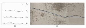
Figure 1. (a) Creating of the fractal curve S, with the curve generator S1 applied to each segment of the curve in each Sk case. (b) Real image of a crack in concrete; the blue lines represent case S0.
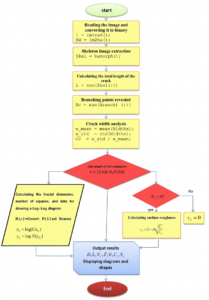
Figure 2. The algorithm applied to analyze crack images in concrete using the Box-Counting method.
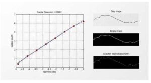
Figure 3. Gray, binary, and skeleton images, and the log-log diagram of the proposed fractal body
The results also appear that the local roughness values Sj are almost constant across the crack path, and the values for the degree of fragmentation F and density are very low. Furthermore, the orientation is nearly horizontal and the shape is centered, which is logical based on our hypothesis of the fractal body. The curve in the log-log diagram of the fragmentary body appears linear, with its points lying almost on a straight line, from which the fractal dimension D is calculated. This corresponds to the assumption that the crack is unbranched and to the value of Nc.
After applying the code, it was possible to observe the difference in the fractional properties of concrete cracks in the different cases studied.
First case: A crack in a wall
(Figure 4.) shows the real and binary (black and white) images of a crack in a wall after filtering; the images of the crack after it has been covered with a grid of squares with their side lengths Ɛ={2,4,8,16,32,64}and only the squares through which the crack passes to count the repetitions. The figure shows that the distribution of the resulting number of squares is similar from scale to scale. Focusing on three fixed areas of the real image, the left-sloping area slanting downwards: at Ɛ=64 one square touches the line, at Ɛ=32 the same area it transforms into three squares, and at Ɛ=16 the same area it transforms into six squares.In the area of the sharp turn in the middle: at Ɛ=64 two adjacent squares, at Ɛ=32 the same relative position, there are 5 squares; at Ɛ=64the same position, there are 10-12 squares. In the area of the wavy section on the right: at Ɛ=64 four intersecting squares, at Ɛ=32 there are 8 squares; at Ɛ=16 the same point, there are 15 squares.Therefore, by tracing the shape of the crack across the different scales of the grid of squares, it can be said that concrete cracks follow a scale-invariant branching pattern.
The following are the results of applying the code to the image of the crack in the wall that appeared in the Command Window:
Fractal Dimension D : 0.9799
Crack Area : 6555 pixels
Crack Length L : 1523 pixels
Number of Branches Nc : 1
Fragmentation F : 0.000657
Avg. Width w_mean : 4.03 pixels
Std Width w_std : 0.73 pixels
Gap Variation Cv : 0.1819
Local Roughness sj per segment :(0.3233, 0.3233, 00.3244, 0.3233, 0.3233, 0.3233, 0.3233, 0.3233, 0.3233, 0.3233)
Crack Density : 0.0042
Orientation : -20.40 degrees
Centroid (x,y) : (491.3 , 449.0)
The results show that the value of the fractal dimension indicates that the crack is almost one-dimensional during a regular propagation phase, exhibiting the behavior of a linear zigzag crack rather than a branching crack or complex surface. The Nc result indicates a single branch, meaning the crack is unbranched, which is consistent with the D value. The crack length (L) is relatively large compared to the area, suggesting an extended rather than localized crack. The propagation occurred over a long distance without fragmentation. Analysis of the area-to-length ratio shows it is very close to the crack width, indicating internal consistency.
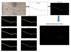
Figure 4. Stages of applying the grid to the crack in the wall after converting the real image to a gray image, then a binary image, and finally a skeleton image, while gradually changing the side length of the grid square Ɛ=64,32,16,8,4,2
This is a strong indicator of the validity of the binary treatment and the skeletonization. The width is also nearly uniform along the crack. The fragmentation degree (F) is very low, indicating no disintegration or splintering of the crack structure. The roughness values are low to medium, indicating a non-perfectly smooth surface, and the density is very low due to the presence of a single crack without secondary cracks.
The Second Case: A Crack In a Beam
Images were taken of a cracked beam (reinforced structural element), and the proposed code was applied to them. (Figure 5.) shows a real image of a crack in a beam; in addition to the images after processing (grey, binary, skeleton); and then the grid was applied to them at different scales.
At large squares , a small number of squares cover the crack path, and the squares contain large gaps, and the crack appears almost straight inside the squares; these shapes reflect the general structure.At medium scales, the crack begins to cut the squares in a zigzag pattern, and the number of squares is almost regular; without any new branching appearing when the scale is reduced. However, at smaller scales, we find that the shape inside each square is similar, and there are no abrupt changes observed in the number of squares; this reflects a stable behavior free from complex branching patterns.
The following are the results of applying the code that appear in the Command Window:
Fractal Dimension D : 0.9952
Crack Area : 4747 pixels
Crack Length L : 937 pixels
Number of Branches Nc : 1
Fragmentation F : 0.001067
Avg. Width w_mean : 4.80 pixels
Std Width w_std : 1.09 pixels
Gap Variation Cv : 0.2265
Local Roughness sj per segment( 0.3199,0.3183,0.3199,0.3183, 0.3199,0.3199,0.3183,0.3199,0.3183,0.3199)
Crack Density : 0.0030
Orientation : 70.17 degrees
Centroid (x,y) : (740.7 , 513.6)
The results show that the value of the fractal dimension is  meaning that the crack is single-path, and that there are no branches in the crack. This is consistent with the Skeleton
image and the value of D. The area-to-length ratio is very close to the width value. This indicates that the calculations are internally consistent, but there ia local fluctuation in the widthwith a low value for the segmentation factor F.Third case: A crack in a column(Figure 6.) shows a real image of a crack in the concrete of a structural column, in addition to the gray, binary and skeleton images, with a grid applied to the extracted image on the exact path of the crack.

Figure 5. Stages of applying the grid to the crack site in the beam after converting the real image to a gray image, then a binary image, and finally a skeleton image, while gradually changing the side length of the grid squareƐ=64,32,16,8,4,2

Figure 6. The actual image of a crack in a column, from which the gray image, the binary image, and the skeleton image were extracted, and a box-counting grid was applied to the crack path with a gradient of squared side lengths Ɛ=64,32,16,8,4,2
We noticed from the real image of the column that the longitudinal crack is relatively wide and continuous, and the edges are irregular. The presence of light and dark areas inside the crack is evidence of a change in depth or illumination. However, the processing has solved this, as the gray image has preserved the general geometric structure. The binary image has isolated the crack and removed the surrounding noise, which is necessary for the Box-Counting to be reliable. The crack in the skeleton image also appears clear. The shape here does not indicate a multi-level hierarchical branching, but only two branches. The grid images show no sudden jumps in the number of squares. This indicates partial self-similarity and good linearity in the Log-Log diagram from which the fractal dimension is taken.It was observed that the shape of the cracks in the reinforced structural elements is more complex, and (Figure 6.) shows the presence of repeating units of squares in the grid across different scales. In the grid images, three types of basic repeating units could be observed:
-Unit A: A small, slanted Z-shaped curve, with individual segments measuring 5-20 pixels. The angle of the curve is small, and this shape appears at all scales.
-Unit B: A short, T-shaped lateral protrusion, approximately 3-10 pixels long. It does not complete as a long branch and appears and disappears with changes in scale. It counts in Box Counting but does not remain in the skeleton image.
-Unit C: A pair of consecutive ∑ bends, consisting of two closely spaced, opposing turns, which are frequently repeated at all scales and are particularly pronounced in more complex regions. It should be noted that these bend units do not repeat in the same magnitude, but they do repeat in the same morphological pattern across different scales (Self-Affine Similarity). Units A and C can be observed to be similar to the fragmentary body shape seen in (Figure 1.).
The following are the results of applying the code that appear in the Command Window:
Fractal Dimension D : 0.9799
Crack Area : 6555 pixels
Crack Length L : 1523 pixels
Number of Branches Nc : 1
Fragmentation F : 0.000657
Avg. Width w_mean : 4.03 pixels
Std Width w_std : 0.73 pixels
Gap Variation Cv : 0.1819
Local Roughness sj per segment:( 0.3233,0.3233,0.3244,0.3233,0.3233, 0.3233,0.3233,0.3244,0.3233,0.3233)
Crack Density : 0.0042
Orientation : -20.40 degrees
Centroid (x,y) : (491.3, 449.0)
We find that the value of the fractal dimension of the crack is linear with a complexity resulting from the zigzag and this is justified by the value of meaning a secondary branch. The values of the length and width are large compared to the previous cases; this is due to the fact that the studied element is a column (a crack resulting from the corrosion of the reinforcing steel), and the width is irregular, but the value of F is very low, implying that the crack is continuous, meaning there is no dissociation into independent sections, with an average oscillation and a uniformly distributed roughness, as the roughness values are close across the sections.Fourth Case: A Crack In a Slab(Figure 7.) shows a real image of a crack in a slab concrete (reinforced structural element) as well as a gray, binary and skeleton images with a grid applied to the extracted image along the crack path at different scales At scale , the crack takes on a simple geometric form as a one-dimensional object without obvious roughness. At , the actual geometry of the crack path begins to emerge, with large curves appearing, but the behavior is still regular. At , this scale seems to fall within the range of true fractal behavior, where squares line up along the perimeter of the crack, and the tortuosity becomes more pronounced. At , the squares follow almost every curve and cover irregular edges, but their number increases regularly. At , the squares begin to reveal small bumps and local variations at the edges. At the highest scale , the squares follow every vibration or local variation and every small change in direction, but there is no large increase in the number of squares . This indicates that the fractal dimension remains close to the value , and therefore the behavior of the squares across scales takes the form of a regular growth in the number of squares without sharp jumps or loss of linearity. This indicates scale consistency across scales. Also, the squares always line up on the path; they do not fill the inner region, so the value of the fractal dimension is very close to 1. We also note the self-similarity of the shape of the square distribution when reducing .
The following are the results of applying the code that appear in the Command Window:
Fractal Dimension D : 1.0358
Crack Area : 10483 pixels
Crack Length L : 1477 pixels
Number of Branches Nc : 2
Fragmentation F : 0.001354
Avg. Width w_mean : 6.43 pixels
Std Width w_std : 1.01 pixels
Gap Variation Cv : 0.1570
Local Roughness sj per segment:( 0.3063,0.3052,0.3063,0.3052,0.3063, 0.3063,0.3052,0.3063,0.3052 0.3063)
Crack Density : 0.0258
Orientation : 4.55 degrees
Centroid (x,y) : (430.7, 285.9
The results show that the value of the fractal dimension is D = 1.0358, meaning that the crack is semi-linear, and this is the largest among the fractal dimension values studied in the previous images. This is also consistent with the fact that the crack consists of two branches without dense branching. The length of the crack is relatively long compared to the area. This is due to the accumulation of the products of the corrosion of the reinforcing steel.
DISCUSSION
Analysis of concrete crack images, after applying a code to process images of damaged structures for different cases (wall, beam, column, slab) using fractal geometry, revealed that these cracks follow a recurring fractal shape. This shape consists of two similar triangles that are axially opposite. With the repetition of this shape several times, the true crack shape that can be observed in the damaged structures is created. The simple fractal body shape was proposed in (Figure 1.), illustrating the fractal crack formation process across three stages. It was shown that the proposed shape can indeed be observed as recurring units in different cracks.
After its presence was confirmed in these cracks, the fractal analysis code was applied, which involves calculating several indicators that can describe the crack state from a fractal perspective.
The most important of these indicators is the fractal dimension, which allows us to measure the complexity of the shape (the crack) (17). Therefore, the code was initially applied to the fractal body proposed in (Figure 1.) and its indicators were calculated. The result showed that the fractal dimension is approximately equal to 1, which corresponds to the formation of a simple, quasi-linear crack and often indicates that there are no branchings in the crack (4). It should be noted that the higher the value of D (above 1), the more complex the crack is and the more urgent it needs maintenance. This is consistent with studies conducted on the fractal dimension (7).
The fractal analysis code was applied to the remaining images, and box-counting grids were applied and displayed only along the crack path to calculate the number of squares that the crack passes through, decreasing the square scale each time. The indices were calculated to ensure that the results were consistent with the proposed fractal body and to characterize the cracks.By tracking the number of squares and their distribution pattern across different scales, starting with the first case of the wall, it became clear that there were repetitions of the distribution pattern with proportionally multiple numbers across the scales, and therefore it can be considered a constant branching pattern across the scales. The calculated indicators also proved to be consistent with the fractal body indicators, showing that the resulting cleft is unbranched, one-dimensional, and proportional to a simple, uncomplicated crack shape.In the second case, that of the beam, an uncomplicated pattern of the crack shape was observed, where the number of squares remained regular across scales, without any sudden jumps in this number, reflecting stability or consistency across scales.However, in the third (column) and fourth (slab) cases, the effect of corrosion on the crack shapeis clearly evident. In the column case, corrosion products accumulate under the concrete cover, forming a more complex crack pattern. The calculated indicators show a higher fractal dimension value compared to the previous cases. This value is consistent with the presence of two crack branches, confirming that the structure is more complex, yet it still maintains regularity. The study of the shape and number of squares reveals three repeating unit shapes across the scales,most of which resemble the proposed fractal shape. It was also observed that the crack width, density, and length are larger.The slab case study shows the largest value for the fractal dimension, which is indeed consistent with the large area of the gap formed in the slab and the volume of the corrosion products. This is the reason for the formation of two crack branches. However, through studies of the number and shape of the squares that define the crack path, their distribution can be considered regular across the scales without any sudden jumps.
The consistency of the shape across scales in all studied images is considered evidence that the crack shape is a fractal shape, as the study showed that the proposed fractal body is consistent with the results obtained from multi-scale Box-Counting tests in terms of the shape of the formed crack and its calculated indices.It turns out that the more complex the crack path is and the larger the area it spreads over, the more the proposed structure of the fractal body has a different dimension and an advanced stage of fragmentation (the images of the column and slab showed that the crack consisted of two branches and was more complex, and the fractal dimension increased compared to the images of the wall and beam and the basic simple fractal body image).
CONCLUSIONS AND RECOMMENDATIONS
In conclusion, using fractal geometry provides a quantitative and objective tool for diagnosing the condition of damaged concrete structures, thus aiding in the assessment of maintenance and reinforcement requirements. Furthermore, combining image processing and fractional analysis constitutes an effective method for evaluating damaged buildings without the need for destructive testing. The consistency of shape across scales in all studied images indicates that the crack shape is fractional. The study demonstrated the agreement of the proposed fractal body with the results obtained from multi-scale box counting tests in terms of the shape of the formed crack and its indices. Therefore, a single cracking indicator is insufficient; the more indicators studied, the more accurate the early prediction of structural element failure or maintenance needs. This study demonstrated the effectiveness of analyzing multiple indicators based on a fractal model. The structural element itself plays a crucial role in the risk of cracking. Load-bearing elements (columns, slabs) are more impactful because any loss of their strength directly affects the building’s overall load-bearing capacity and corrosion. It is recommended to expand the database by applying the methodology to a larger number of images and a variety of structures (bridges, columns, slabs) to generalize the results. Multiscale analysis can also be applied instead of singular fractal dimension to obtain a more accurate characterization of the crack network, in addition to select different fractal structures that closely resemble cracks for use in an early warning system for building and bridge maintenance, and to increase the number of indicators studied for crack characterization.


![Study the optimal Conditions for the Determination of Zinc (II) in Aqueous Solutions Using Organic Reagent 4-Hydroxy – 3 -[[hydroxy phenyl) methylene] amino] benzene sulphonic acid II by Spectrophotometric method](https://journal.hcsr.gov.sy/wp-content/uploads/2026/05/صورة-مقال-عمار-اسماعيل.png)